
Medical Services

Refractive Surgery Consultation
Dr. Marlatt has been co-managing patients undergoing refractive surgery for the last 8 years. He will be able to discuss all of your options including LASIK, PRK and refractive lens exchange.
BOOK NOW
Routine Comprehensive Assessment
The standard Routine Comprehensive Assessment consists of a complete eye exam excluding imaging. Refraction using digital phoropter is included. For those who wear spectacles, a prescription measurement is provided and the UV (Ultra Violet) protection and blue light penetration of your current lenses will be measured.
A complete general ocular health assessment will be performed. Vision acuity, pupillary function, colour vision, peripheral vision, ocular motility and alignment will all be evaluated and assessed. If determined necessary, this exam may be paired with digital retinal photos or an Optical Coherence Topography (OCT) scan to provide a more extensive look at your optical health.

Premium Comprehensive Assessment
The Premium Comprehensive Assessment will provide the most complete ancillary examination available. This assessment includes the components of the routine comprehensive assessment as described above, and includes digital retinal photos, optical coherence topography of the retina and optic nerve head, pachymetry (corneal thickness), mapping of the cornea, a scan of the anterior chamber, automated perimetry and meibography.
BOOK NOW
Urgent Eye Care
Serious and acute eye conditions or injuries can occur unexpectedly and at any time. A sudden significant decrease in vision, persistent flashes continuing for more than an hour, an onset of floaters, and double vision are all indications of eye disease or trauma and may be critical to your ocular health. Many of these ailments may or may not be accompanied by pain. If any of these occur, consider it urgent and seek the service of an eye care professional immediately or report to the nearest emergency department.
BOOK NOW
Contact Lens Fitting
Contact lenses for all vision correction needs are provided with a personalized fitting. Lenses for distant or near vision, astigmatism, multifocal, mono-vision, and rigid contact lenses are available. Dr. Marlatt has extensive experience helping patients with specialty contact lenses, including scleral.
BOOK NOW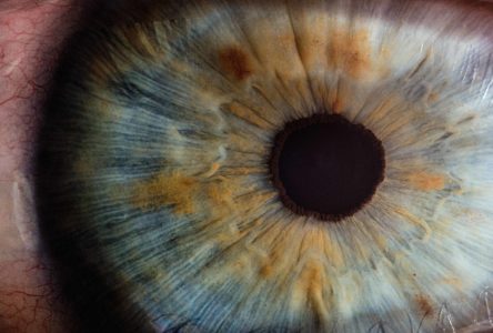
Digital Retina Imaging
Fundus photos not only provide insight into ocular health, but can provide much information about overall health. A twelve megapixel camera utilizes an intricate microscope and low intensity flash to provide a view of the posterior chamber of the eye. The result is an image with high resolution and contrast, showing retinal vascular, macula and optic nerve head appearance as well as the choroid, which cannot be seen by other ocular exam methods. Retinal imaging creates a baseline which allows for documenting the status and changes which may occur otherwise undetected.
BOOK NOW
Optical Coherence Topography (OCT)
This non-invasive imaging technique generates 3μm high-resolution images by scanning 68000 points (A-scans) per second. This allows the measurement of layers of distinctive anatomical structures in both the anterior and posterior sections of the eye. This technology may be used for screening or monitoring pathology of the cornea, retina and optic nerve. Some structures can be visualized in 3D to help with diagnosis and management of eye disease.
BOOK NOW
Perimetry
Visual field analysis is a systematic measurement used to monitor central and peripheral vision function. Utilizing a variety of threshold algorithms and screening techniques, these measurements are significant in the management of macular disease and glaucoma progression. The the Esterman Binocular Driver’s Test; 30-2, 24-2 and 10-2, and blue on yellow in Golmann Standard are used.
BOOK NOW
Meibography
The Meibomian(my-bo-me-an) glands naturally secrete a stabilizing lipid layer of tear film to keep eyes moist, lubricated and healthy. A specialized imaging study, developed exclusively for the purpose of visualizing the morphology of meibomian glands, is used to determine the Tear Breakup Time (TBUT). This reading is beneficial in determining and understanding ocular surface diseases and managing Dry Eye Syndrome.
BOOK NOW
Glaucoma Management
Dr. Marlatt has access to the latest technology to diagnose and manage primary open angle glaucoma. He also has an exceptional network of specialist who help manage those in need of additional care.
BOOK NOW